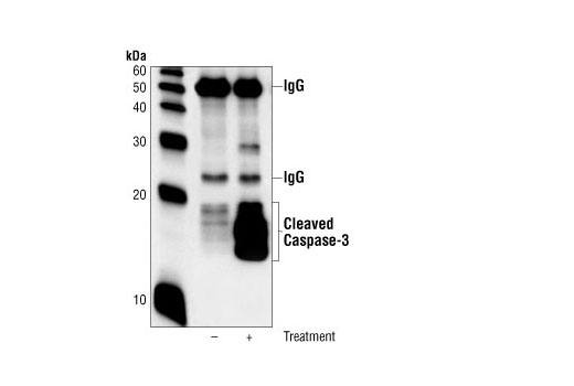
Cleaved Caspase 3 Western Blot
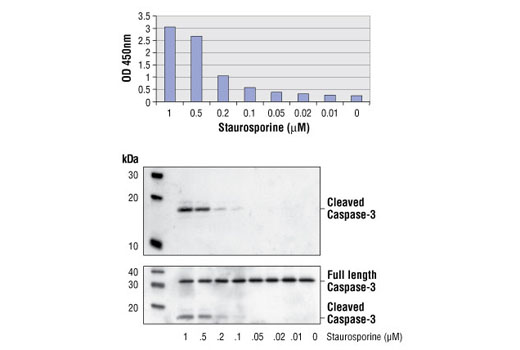
Pathscan Cleaved Caspase 3 Asp175 Sandwich Elisa Kit
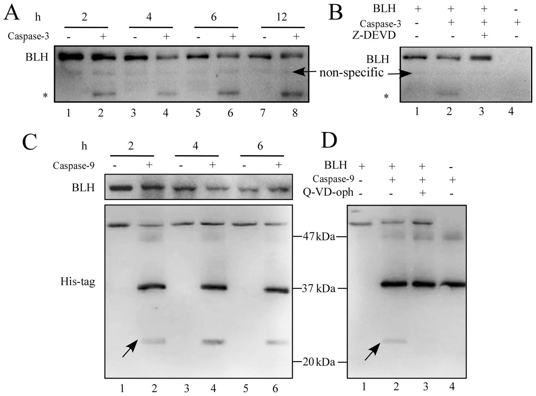
Cleavage Of Bleomycin Hydrolase By Caspase 3 During Apoptosis
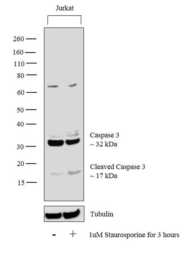
Caspase 3 Antibody Ma1

Nanoparticle Delivery Of Artesunate Enhances The Anti Tumor Efficiency By Activating Mitochondria Mediated Cell Apoptosis Abstract Europe Pmc

Recombinant Human Cleaved Caspase 3 Protein Ab Abcam

Western Blot Analysis Of l 2 Cleaved Caspase 3 And Cytc Protein Download Scientific Diagram
Caspase-3, Apopain, Cysteine protease CPP32, Protein Yama, SREBP cleavage activity 1, CASP3_HUMAN.
Cleaved caspase 3 western blot. Western blot analysis confirmed that the caspase-3 inhibitor completely blocked the cleavage of Par-4 in WT caspase-3–Myc-transfected MCF-7 cells treated with cisplatin (Fig. Apigenin is a flavonoid nutrient and possesses antineoplastic actions. A8K5M2, D3DP53, P, Q96AN1, Q96KP2.
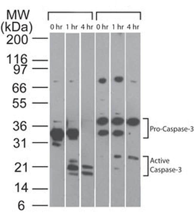
And I find some researchers used Caspase-3 activity assay kit in literature. A large number of irradiated colorectal tumor cells (feeder cells) were then co-cultured with a small number of luciferase-labeled living colorectal tumor. Caspase-3 Antibody (31A1067) - (Pro and Active) NB100- - Image of Caspase-3 Antibody (31A1067) - (Pro and Active).
Western Blot Using Caspase-3 (3G2) Mouse mAb from Cell Signaling Technology (1) Write a Review. Caspase-1 Antibody (D-3) is a high quality monoclonal caspase-1 antibody (also designated CASP1 antibody) suitable for the detection of the caspase-1 protein of mouse, rat and human origin. Cleavage and activation of pro caspase-3 is catalyzed by caspase-8, caspase-9, and granzyme B to generate the active heterodimer of caspase-3 subunits (1).
Caspase-3 activation is detected in Western blots by the presence of cleavage fragments. The Apoptosis western blot cocktail (ab) is designed to study the induction of apoptosis in response to various stimuli. Human COLO cell lysate.
The cleaved caspase-3 (CC3) in irradiated tumor cells and HMGB1 in the supernatant of irradiated tumor cells were detected by Western blot. The immunoblot was a representative of three independent experiments. Effect of ZVAD on caspase 3 activity.
Human COLO cell lysate treated with the immunizing peptide. AA6, monoclonal ChIP, IHC, IP, WB pig, rat, human, mouse, rat, rabbit, chicken, rabbit, pig, guinea. Caspase 3, also named as CPP32, SCA-1 and Apopain, belongs to the peptidase C14A family.
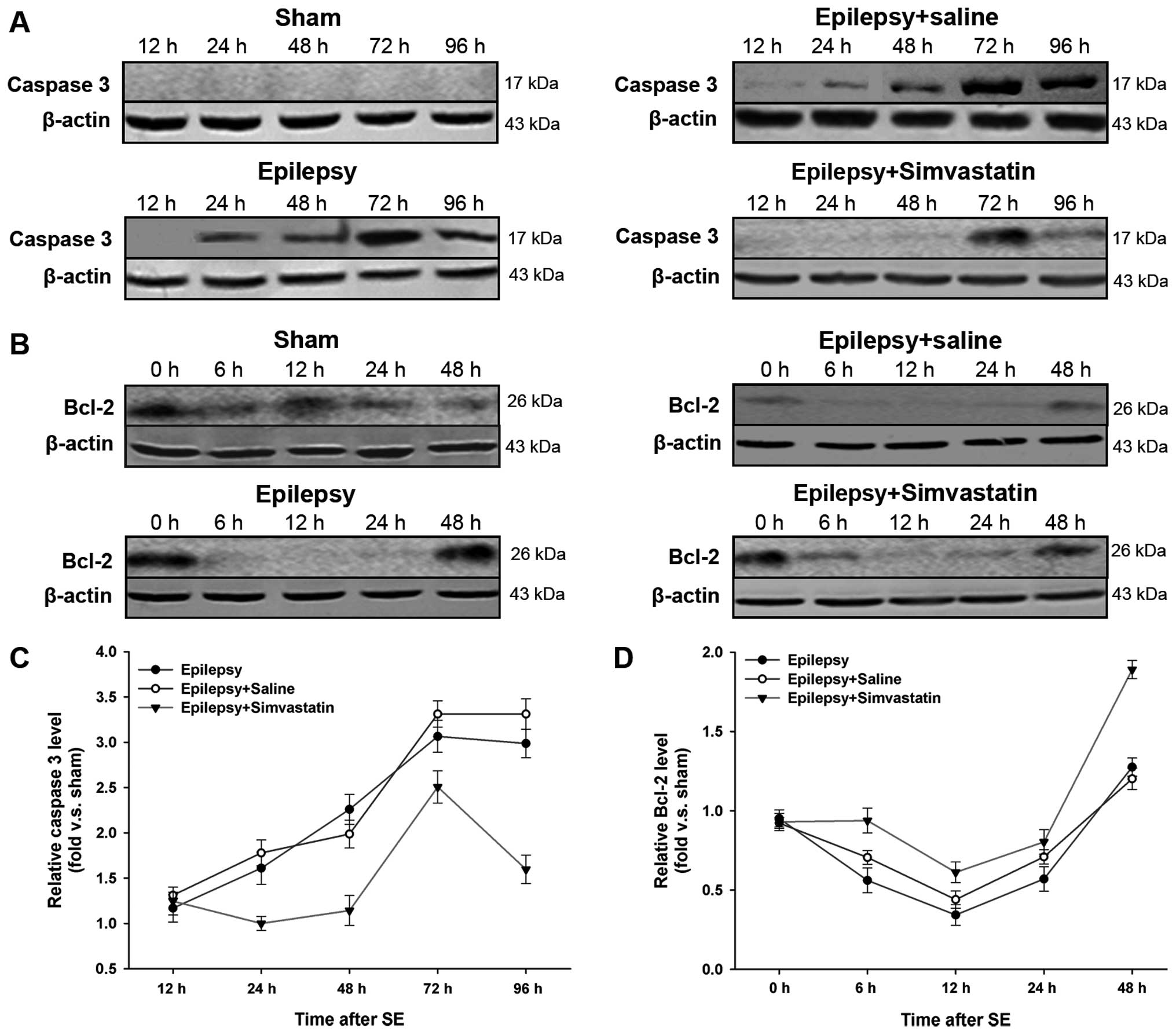
(A) Fluorescence staining images of PC12 cells treated with miR-181c mimic or inhibitor. One of the key mediators of apoptosis is the thiol protease caspase 3. (C) Representative western blots of l-2, Bax, and cleaved caspase-3.
Western blot analysis of the effect of SAM-486A, DFMO and NOHA on the activation and proteolytic cleavage of caspase-3 in MDA-MB-468 cells. The lane on the right is blocked with the synthesized peptide. DFFA is the substrate for caspase-3 and triggers DNA fragmentation during apoptosis.
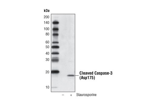
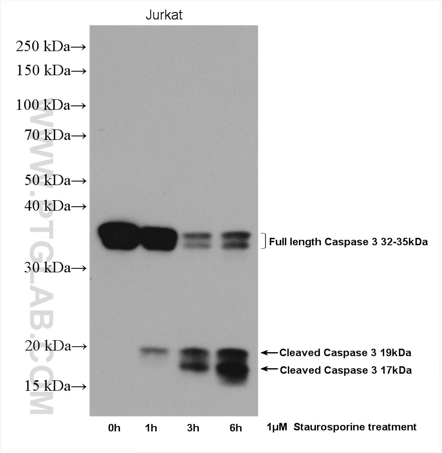
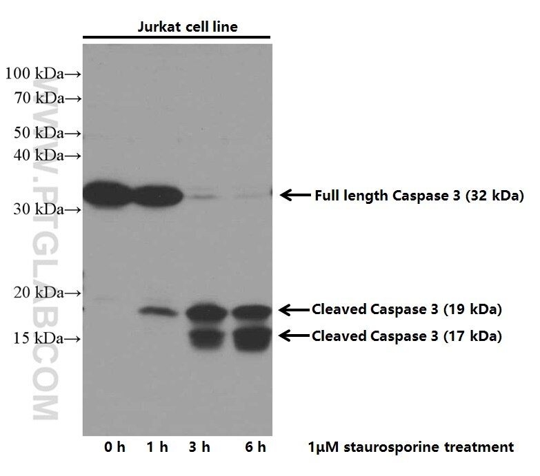
"Recombinant Newcastle disease virus (rL-RVG) triggers autophagy and apoptosis in gastric carcinoma cells by inducing ER stress.". Sagulenko V, Vitak N, Vajjhala P, Vince J, Stacey K. Western blot shows lysates of Jurkat human acute T cell leukemia cell line untreated (-) or treated (+) with 1 µM staurosporine (STS) for 3 hours.
Cell proliferation inhibition, induction of apoptosis, cell cycle arrest and in…. Search results for cleaved caspase 3 at Sigma-Aldrich. Caspase 3 is one of the executioner caspases activated by proteolytic cleavage during apoptosis.
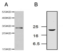
DFF becomes activated when DFFA is cleaved by caspase-3. B , western blotting analysis of Jurkat cell lysates, from cells treated with camptothecin, probed with Rabbit Anti-Caspase-3 (active) Antibody ( AHP2286 ), which detects a band of approximately 17 kDa. Cell Signaling Technology's Cleaved Caspase-3 (D175) Antibody is a polyclonal antibody that cross reacts with human, rat, and mouse caspase-3.
The antibody detects both pro (full-length) and active (cleaved) protein. Caspase 3 is cleaved at Asp28/Ser29 and Asp175/Ser176. The cleaved fragments of DFFA dissociate from DFFB, the active component of DFF.
TT genotype of CASP3 rs polymorphisms showed risk in CAD. Proteins were transferred to a membrane and probed with a Caspase 3 (active/pro) Monoclonal Antibody (31A1067) (Product # MA1-) at a dilution of 5 µg/mL. Validation of Anti-CSPα, SNAP25, Tyrosine Hydroxylase, Ubiquitin, Cleaved Caspase 3, and pSer PKC Motif Antibodies for Utilization in Western Blotting Acta Histochem Cytochem.
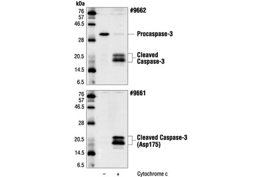
Western blot analysis of extracts from C6 (rat), NIH/3T3 (mouse), and Jurkat (human) cells, untreated or treated with staurosporine #9953 (1uM, 3hrs) or etoposide #20 (25uM, 5hrs) as indicated, using Cleaved Caspase-3 (Asp175) (5A1E) Rabbit mAb. Activation of the extrinsic or intrinsic pathway leads to the cleavage of caspase-3 (or caspase-7), which leads to the commitment to apoptotic death, and is thus considered a reliable apoptosis marker. Western blot analysis of cytochrome c and cleaved caspase-3 protein.
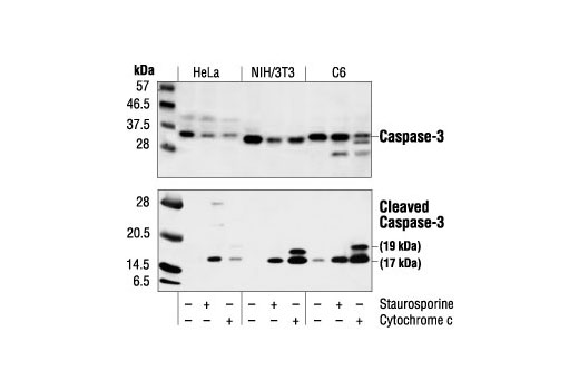
Western blot analysis of lysates from 293 cells, treated with Etoposide 25uM 60', using Caspase 3 (Cleaved-Asp175) Antibody. Western blot analysis of extracts from HeLa, NIH/3T3 and C6 cells untreated, staurosporine-treated (3hrs, 1 µM in vivo) or cytochrome c-treated (1hr, 0.25 mg/ml in vitro), using Caspase-3 Antibody #9662 (upper) or Cleaved Caspase-3 (Asp175) Antibody (lower). Caspase 3 activity was determined by Western blot in control cells and cells treated with Cas III-ia and Cas III-ia + 50μΜ ZVAD for 24 h.
Bu, Xuefeng, et al. Concurrently, pretreatment with the caspase-3 inhibitor significantly reduced the number of cells entering apoptosis and inhibited the activation of caspase. In this investigation, caspase 3 mRNA and protein expression in breast cancer was examined.
Authors Toshihiko Shirafuji 1. Cleaved Caspase-3 (Asp175) Western Detection Kit offers an efficient way of detecting caspase-3 processing and activation by Western blotting. Whole cell protein from Jurkat cells treated with and without 2 uM staurosporine as indicated was separated on a 4-15% gel by SDS-PAGE, transferred to 0.2 um PVDF membrane and blocked in 5% non-fat milk in TBST.
We resolved recombinant activated caspase-3 by 15% SDS–PAGE and transferred to a NC membrane, and the blotted membrane was incubated with different. Rabbit Anti-Caspase 3 Polyclonal Antibody (SPC-1319) at 1:1000. Western blot analysis of Human COLO cell lysates showing detection of ~17kDa Caspase 3 protein using Rabbit Anti-Caspase 3 Polyclonal Antibody (SPC-1319).
Caspase-1 Antibody (D-3) is available as both the non-conjugated anti-caspase-1 antibody form, as well as multiple conjugated forms of anti-caspase-1 antibody, including agarose, HRP, PE, FITC and multiple. DNA fragmentation factor (DFF) is a heterodimeric protein of 40-kD (DFFB) and 45-kD (DFFA) subunits. Anti-Caspase 3 Antibody, active (cleaved) form detects level of Caspase 3 and has been published and validated for use in Immunofluorescence (IF), Immunohistochemistry (IHC), and Western Blot (WB).
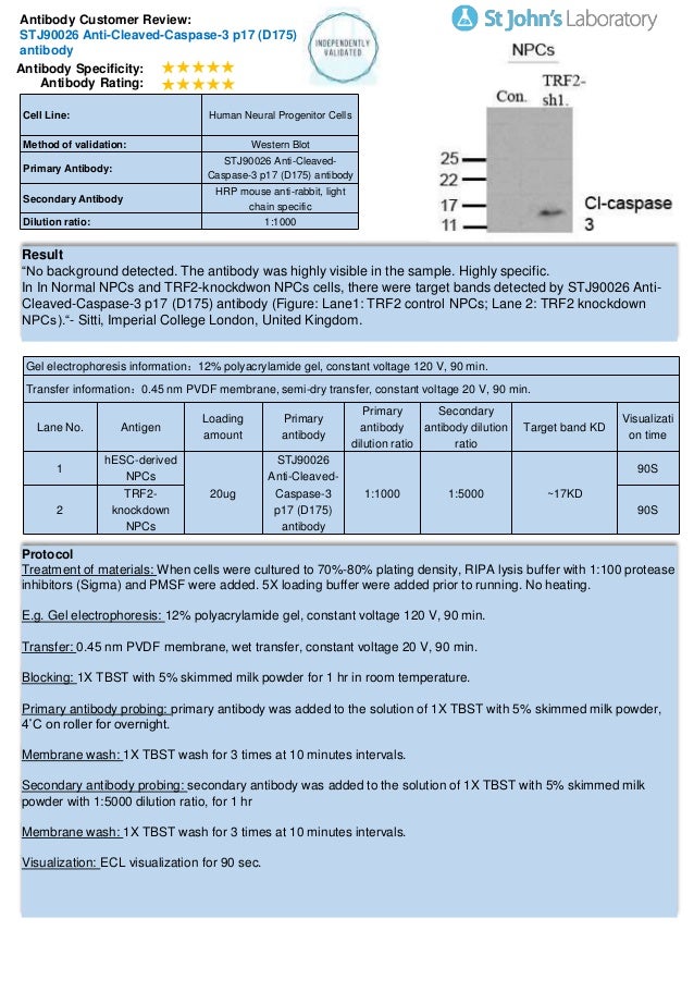
The increased representation of cleaved caspase-3 in KIAA1199 knockdown cells compared to the control cells is qualitatively shown in MDA-MB-231 (left panel) and Hs578T (right panel) cells. The absence of signal in the CASP3 knock-out HCT116 cells confirms specificity of the antibody for CASP3. Western Blot analysis of KB cells using Cleaved-Caspase-3 p12 (D175) Polyclonal Antibody.
Similarly, procaspase-3 (~32 kD) is processed to the p/p17 bands, the latter representing catalytically active caspase-3 (5-10;. Horizon offers western blot analysis of cleaved caspase-3 and full length and cleaved PARP to study the induction of apoptosis. A8K5M2, D3DP53, P, Q96AN1, Q96KP2.
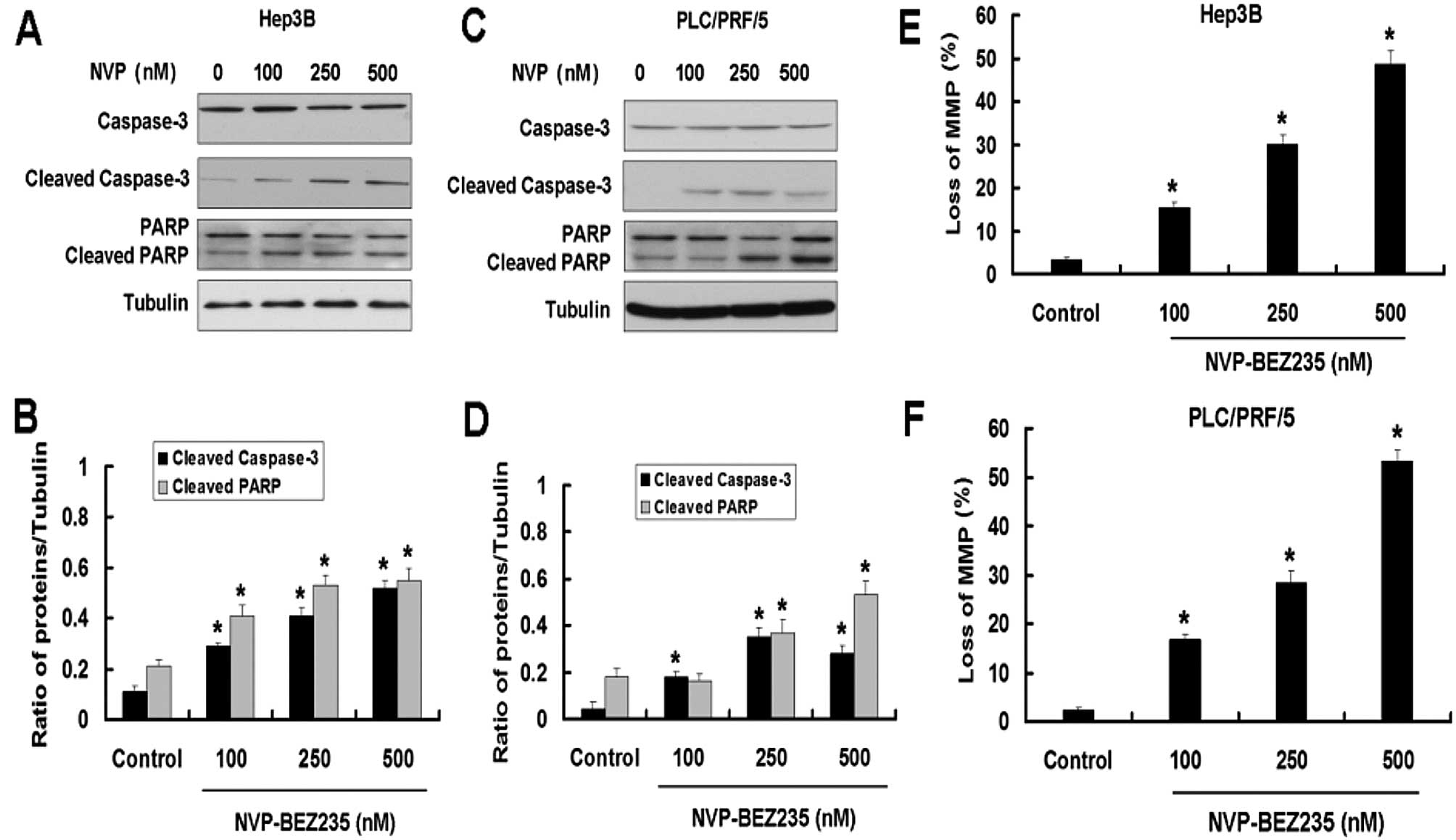
Cleaved Caspase 3 is a well-known marker for cells undergoing apoptosis in the caspase-dependent pathway. Cleaved caspase-3 and full length caspase-3 protein expression was determined via western blotting. Caspase 3 was measured at the mRNA level using reverse transcription-PCR and at the protein level using both Western blotting and activity assays.
Some customers have used this antibody successfully in IHC-P however our latest tests were unsuccessful and therefore we can no longer guarantee this application. I have detected the apoptosis in the organ of cochlea by Western blot using a cleaved caspase-3 antibody. Western Blot analysis of Caspase 3 (active/pro) was performed by loading Jurkat cell lysates treated with and without 2 uM staurosporine.
Cells (2×10 6 ) were treated with SAM-486A and DFMO, either alone or in combination, for days 3 and 6 and with NOHA (1 mM) until day 2 (48 h), lysed in lysis buffer, and mg of cell lysate were run on 10. Our results showed that TG treatment markedly increased the expression of CHOP, cleaved-caspase-12 and cleaved-caspase-3, but the alterations were significantly inhibited by Activin A treatment (B). It does not recognize endogenous levels of full length caspase-3 or other caspases.
In the HG group, cleaved caspase-3/full length caspase-3 protein levels were significantly increased. This antibody reacts with Human, mouse, rat samples. Data presented as the mean ± standard deviation of three independent experiments.
β-Actin was used as the internal control. Active caspase-3 detection by western blotting. Detection of Human and Mouse Cleaved Caspase‑3 (Asp175) by Western Blot.
Caspases 6, 7 and 9), as well as relevant targets in the cells (e.g. Monoclonal antibody is produced by immunizing animals with a synthetic peptide corresponding to amino-terminal residues adjacent to Asp175 of human caspase-3. Western blot shows lysates of Jurkat human acute T cell leukemia cell line and DA3 mouse myeloma cell line untreated (-) or treated (+) with 1 µM staurosporine (STS) for the indicated times.
Polyclonal antibodies are produced by immunizing animals with a synthetic peptide corresponding to amino-terminal residues adjacent to (Asp175) in human caspase-3. Cleaved caspase-3 and caspase-3/8/9 could be biomarkers for tumorigenesis in oral tongue squamous cell carcinoma patients. At the onset of apoptosis it proteolytically cleaves poly (ADP-ribose) polymerase (PARP) at a '216-Asp-|-Gly-217' bond.
The protein levels of CHOP, cleaved-caspase-12 and cleaved-caspase-3 were investigated by the western blot assay. PVDF membrane was probed with 1 µg/mL of Human Caspase-3 Monoclonal Antibody (Catalog # MA07), followed by HRP-conjugated. The active Caspase 3 proteolytically cleaves and activates other caspases (e.g.
It can be used for western blotting, immunocytochemistry, and immunohistochemistry. Epub 17 Dec 21. - Find MSDS or SDS, a COA, data sheets and more information.
Caspase-3 is expressed in cells as an inactive precursor from which the p17 and p11 subunits of the mature caspase-3 are proteolytically generated during apoptosis. Caspase-3, Apopain, Cysteine protease CPP32, Protein Yama, SREBP cleavage activity 1, CASP3_HUMAN. Used caspase-3 antibodies to monitor the induction of apoptosis and caspase-3 cleavage and activation through western blotting (2).
5 C and D). Traditional Western Blot Results MSDLY0001 and MSDLY0002 whole cell lysates ( µg each) were analyzed by Western Blot with an antibody recognizing both full length and cleaved Caspase-3. Variations in levels of Caspase 3 have been reported in cells of short-lived nature and those with a longer life cycle.
At 48 h after transfection, the cells were harvested and processed for western blotting. This caspase is responsible for the majority of proteolysis during apoptosis, and detection of cleaved caspase-3 is therefore considered a reliable marker for cells that are dying, or have died by apoptosis. Western blot analysis of extracts from HCT116 cells (lane 1) or CASP3 knock-out cells (lane 2) using Caspase-3 Antibody #9662 (upper), and α-Actinin (D6F6) XP® Rabbit mAb #6487 (lower).
MSD MULTI-ARRAY Cleaved Caspase-3 Assay 0.3 0.6 1.25 2.5 5 10 0 10 30 40 50 60 positive/negative ratio μ g lysate Full Length Cleaved kD MSDLY 0001. Caspase-1 Is an Apical Caspase Leading to Caspase-3 Cleavage in the AIM2 Inflammasome Response, Independent of Caspase-8. This protocol outlines the quantification of apoptosis by flow cytometric detection of cleaved caspase-3.
CASP3 rs creates a new exon splicing enhancer. Because Western blot processing of membranes may lead to protein loss, we tested whether the glutaraldehyde fixation of blotted membranes could improve sensitivity of caspase-3 immunodetection. A , Rabbit Anti-Caspase-3 Antibody detects a band of approximately 32 kDa in Jurkat cell lysate under reducing conditions ( AHP2717 ).
(D) Western blot assay of Bax, l-2, and caspase-3 staining in PC12 cells, including statistics for relative. Caspase 3/p17/p19 antibody Mouse Monoclonal from Proteintech validated in Western Blot (WB), Immunohistochemistry (IHC), Immunofluorescence (IF), Enzyme-linked Immunosorbent Assay (ELISA) applications. The kit contains enough primary and secondary antibodies to perform 10 Western mini blots, as well as a set of pre-stained and biotinylated markers, cell lysates and LumiGLO® reagent.
(B) Cell viability of transfected PC12 cells, as assessed by MTT assay. The caspase-3 precursor is first cleaved at Asp175-Ser176 to produce the p11 subunit and the p peptide. Product Pathways - Apoptosis Cleaved Caspase-3 (Asp175) Antibody #9661 pathway application references companion products datasheet PDF MSDS PDF protocols PhosphoSitePlus® protein, site, and accession data:.
Caspase 3 is involved in the activation cascade of caspases responsible for apoptosis execution. Detection of Human Precursor Caspase‑3 and p18 Subunit by Western Blot. Ab recognizes a cleaved form of Caspase 3 (~17 kDa) after apoptosis has been induced in wildtype cells and not Caspase 3 knockout cells.

Fig 2 Coexistence Of High Levels Of Apoptotic Signaling And Inhibitor Of Apoptosis Proteins In Human Tumor Cells Cancer Research
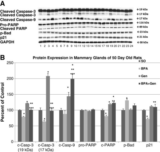
Protein Expressions In Pnd50 Rats Western Blot Analysi Open I
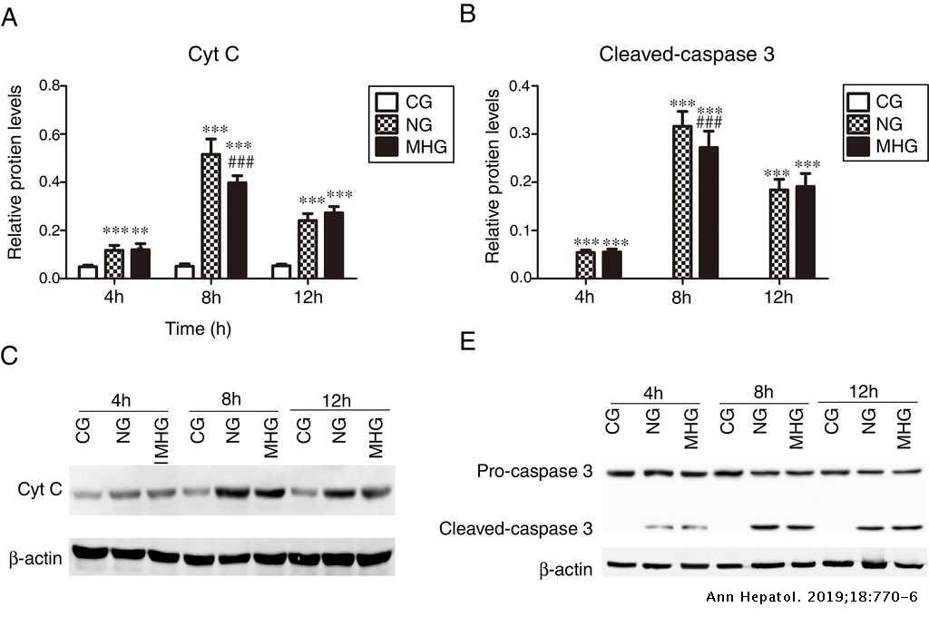
Protective Effects Of Mild Hypothermia Against Hepatic Injury In Rats With Acute Liver Failure Annals Of Hepatology
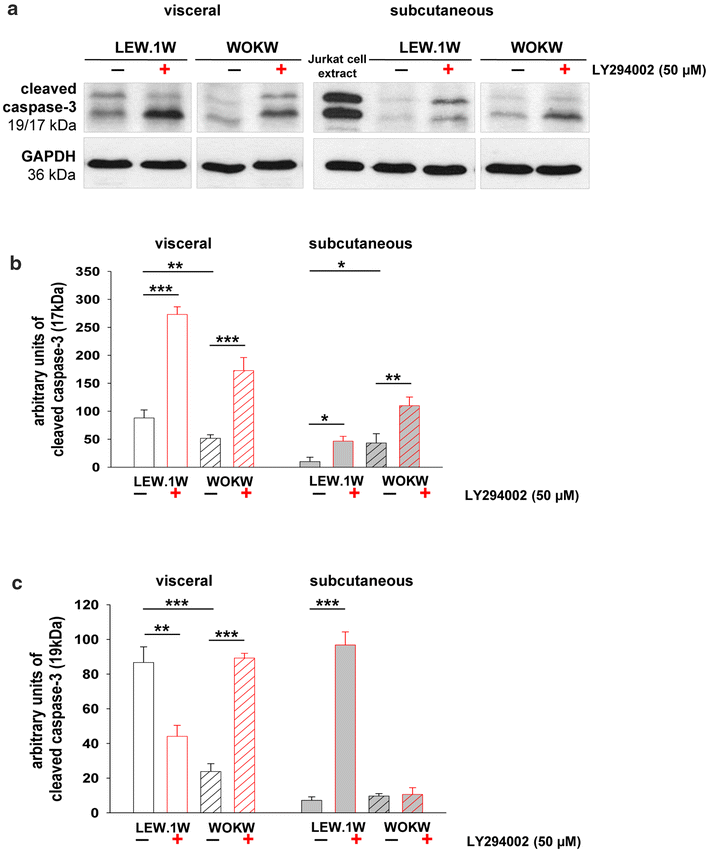
Up Regulated Autophagy As A Protective Factor In Adipose Tissue Of Wokw Rats With Metabolic Syndrome Diabetology Metabolic Syndrome Full Text
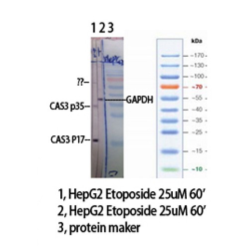
Anti Casp3 Caspase 3 Antibody Rabbit Anti Human Polyclonal Wb Lsbio
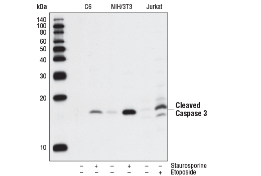
Cleaved Caspase 3 Asp175 5a1e Rabbit Mab
Cleaved Caspase 3 Asp175 5a1e Rabbit Mab Biotinylated

Prostate Apoptosis Response 4 Par 4 A Novel Substrate Of Caspase 3 During Apoptosis Activation Molecular And Cellular Biology
Pro Caspase 3 Antibody Ma1
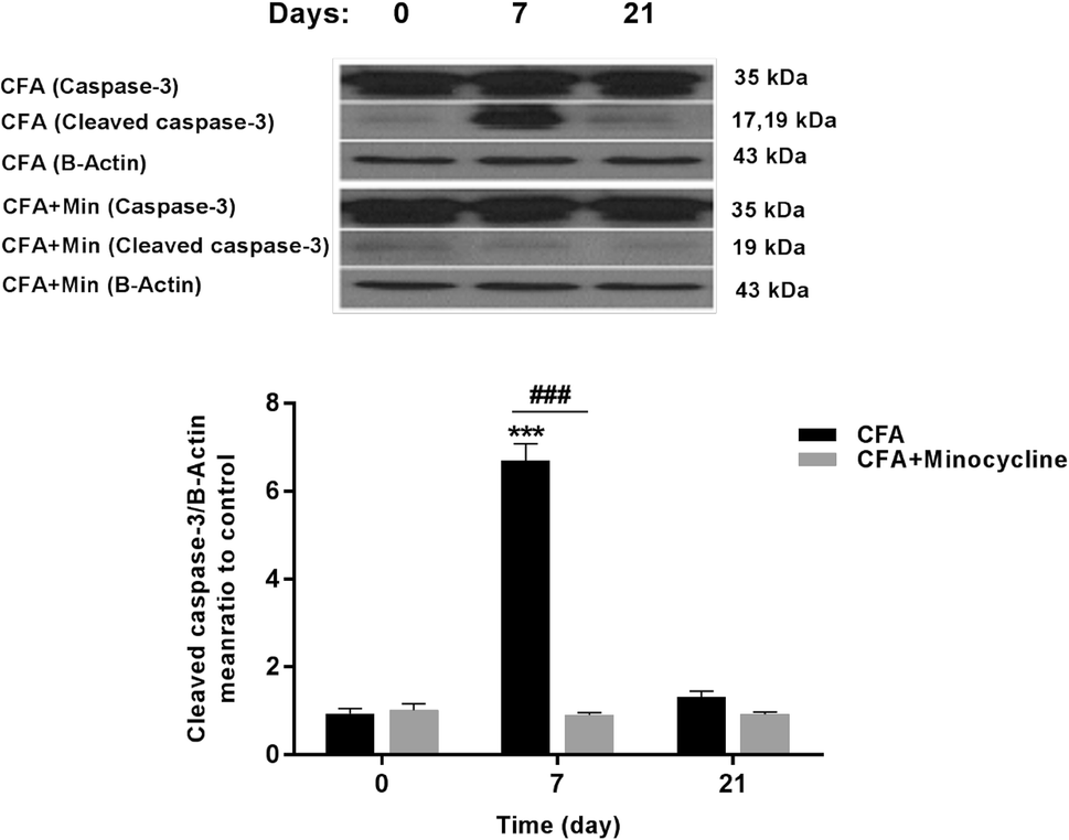
Figure 9 Microglial Induced Apoptosis Is Potentially Responsible For Hyperalgesia Variations During Cfa Induced Inflammation Springerlink

A Caspase 3 Death Switch In Colorectal Cancer Cells For Induced And Synchronous Tumor Apoptosis In Vitro And In Vivo Facilitates The Development Of Minimally Invasive Cell Death Biomarkers Cell Death Disease

View Image

View Image

Cleaved Caspase 3 Asp175 5a1e Rabbit Mab Cell Signaling Technology Inc Bioz Ratings For Life Science Research
Dual Pi3k Mtor Inhibitor Nvp Bez235 Induced Apoptosis Of Hepatocellular Carcinoma Cell Lines Is Enhanced By Inhibitors Of Autophagy
-Western-Blot-NB100-56113-img0006.jpg)
Caspase 3 Antibody Active Cleaved Nb100 Novus Biologicals

17 123
Onlinelibrary Wiley Com Doi Pdf 10 1002 Ijc

Western Blot Analysis Of Pcna Ki 67 Cleaved Parp Cleaved Caspase 3 And Nf Kb In Pc 3 Tumor Tissues Samples

Caspase 3 Antibody Unconjugated Cleaved Asp175 Mab5 Novus Biologicals
Plos One Dissecting The Mechanisms Of Doxorubicin And Oxidative Stress Induced Cytotoxicity The Involvement Of Actin Cytoskeleton And Rock1

Caspase Activation And Neuroprotection In Caspase 3 Deficient Mice After In Vivo Cerebral Ischemia And In Vitro Oxygen Glucose Deprivation Pnas
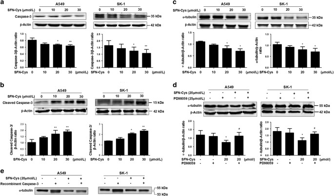
Sulforaphane Cysteine Induced Apoptosis Via Phosphorylated Erk1 2 Mediated Maspin Pathway In Human Non Small Cell Lung Cancer Cells Cell Death Discovery
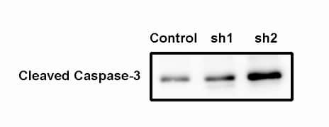
Human Mouse Cleaved Caspase 3 Asp175 Antibody Mab5 R D Systems
Q Tbn 3aand9gcs6tdvllqoujn1bzlh Zbsdwi5movww Rljs8zo2gvzlxl1xdtn Usqp Cau

Cleaved Caspase 3 Asp175 P17 Antibody Af7022 Affinity Biosciences

Caspase 3 Antibody 31a1067 Scbt Santa Cruz Biotechnology

Recombinant Anti Cleaved Caspase 3 Antibody Epr Ab Abcam

Caspase 3 Activation And Increased Procollagen Type I In Irradiated Hearts

Apoptosis Western Blot Cocktail Pro P17 Caspase 3 Cleaved Parp1 Muscle Actin Ab

Efficient Apoptosis Requires Feedback Amplification Of Upstream Apoptotic Signals By Effector Caspase 3 Or 7 Science Advances
Pi 1840 Induces The Apoptosis Of Os Cells A Western Blot Analyses Of Download Scientific Diagram
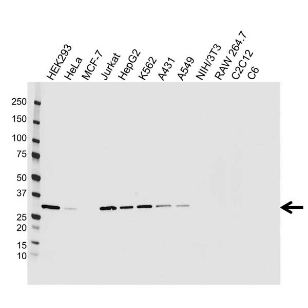
Anti Caspase 3 Antibody Precisionab Monoclonal Antibody Bio Rad

A Western Blotting Analysis For Cyto C Prohibitin Cleaved Caspase 3 Download Scientific Diagram

Recombinant Human Cleaved Caspase 3 Protein Ab Abcam

Efficient Apoptosis Requires Feedback Amplification Of Upstream Apoptotic Signals By Effector Caspase 3 Or 7 Science Advances

Constitutive Activation Of Caspase 3 And Poly Adp Ribose Polymerase Cleavage In Fanconi Anemia Cells Molecular Cancer Research

Human Mouse Caspase 3 Antibody Af 605 Na R D Systems

A Western Blot Analysis Of Bax And Cleaved Caspase 8 Caspase 9 And Download Scientific Diagram
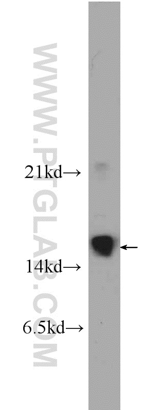
Cleaved Caspase 3 Antibody 1 Ap Proteintech

Fig 6 Molecular And Cellular Biology

View Image

View Image
---(Pro-and-Active)-Western-Blot-NB100-56708-img0031.jpg)
Caspase 3 Antibody 31a1067 Pro And Active Nb100 Novus Biologicals
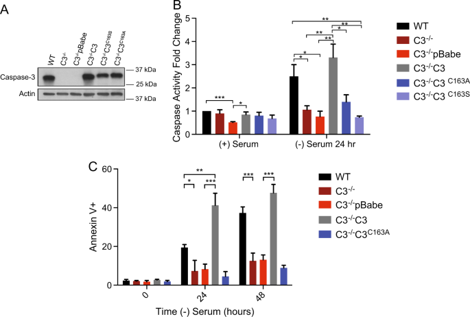
The Prodomain Of Caspase 3 Regulates Its Own Removal And Caspase Activation Cell Death Discovery
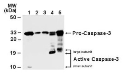
Vanquish Oncology Procaspase 3 Activation Factor To Selectively Induce Apoptosis Cancer Biology

Recombinant Anti Cleaved Caspase 7 Antibody Epr 25 Bsa And Azide Free Ko Tested Ab

Analysis Of Apoptotic Pathways By Western Blot
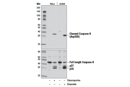
Cleaved Caspase 9 Asp330 D2d4 Rabbit Mab

Signalslide Cleaved Caspase 3 Asp175 Ihc Controls 8104
Plos One Adiponectin Protects Rat Myocardium Against Chronic Intermittent Hypoxia Induced Injury Via Inhibition Of Endoplasmic Reticulum Stress

Liver Wrapping Nitric Oxide Releasing Nanofiber Downregulates Cleaved Caspase 3 And Bax Expression On Rat Hepatic Ischemia Reperfusion Injury Sciencedirect

Apoptosis Antibody Kit

Anti Caspase 3 Antibody Products Biocompare
Plos One Activation Of Chymotrypsin Like Activity Of The Proteasome During Ischemia Induces Myocardial Dysfunction And Death

Nb 22 0027 0ul Anti Cleaved Caspase 3 P17 D175 Antibody Clinisciences
Q Tbn 3aand9gctitevhy I8yzbyi Xau5yhfnecmvzzmpjm75qyksk7adcau Tb Usqp Cau
Q Tbn 3aand9gcqmhvn9aptqzakjfucjltj4ihfpaitflk5egjwaworofq3pxqes Usqp Cau
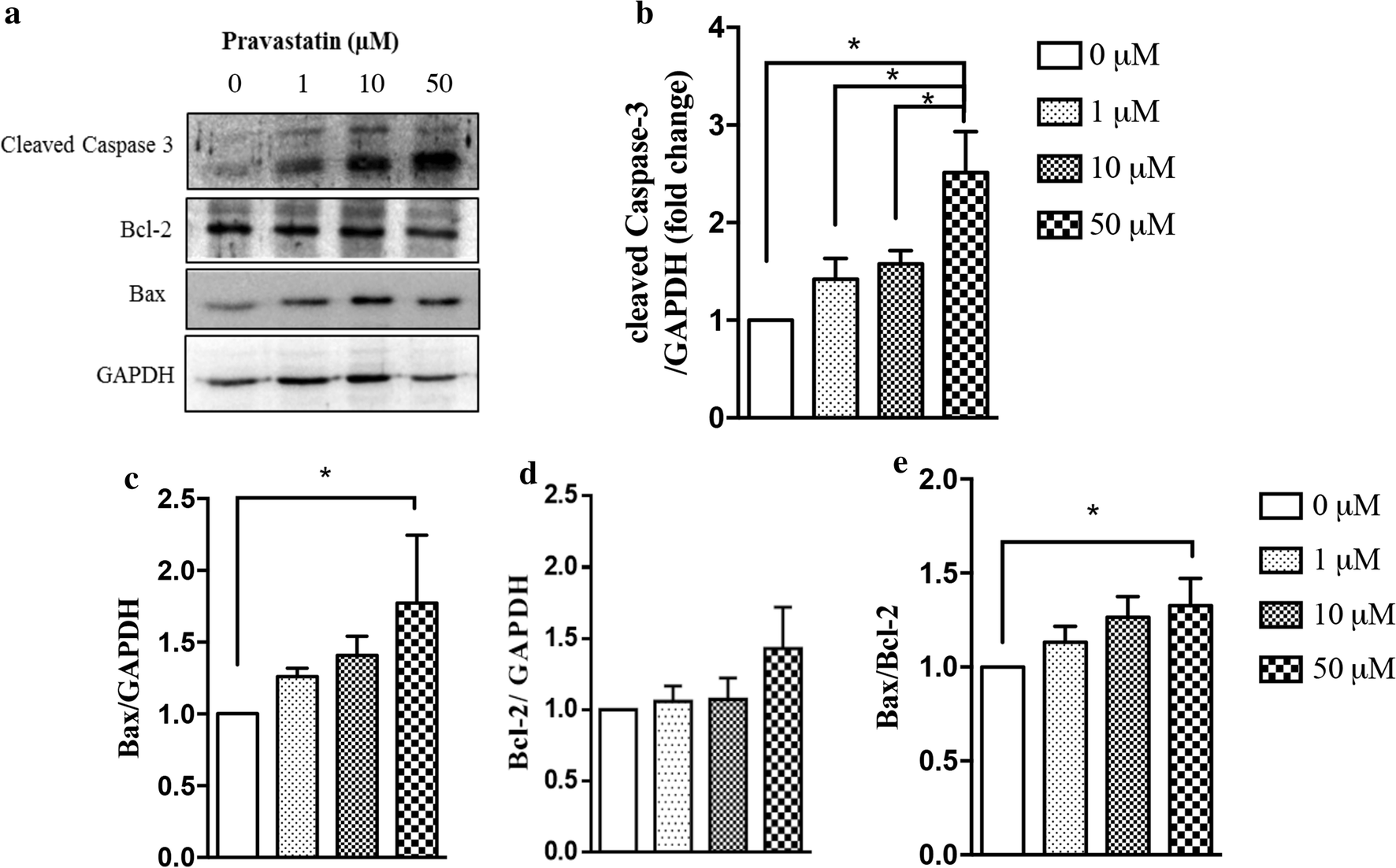
Diabetogenic Effect Of Pravastatin Is Associated With Insulin Resistance And Myotoxicity In Hypercholesterolemic Mice Journal Of Translational Medicine Full Text
Cleaved Caspase 3 Asp175 Antibody
Apoptosis Analysis By Western Blotting Bio Rad

A Representative Western Blot For Cleaved Caspase 3 And Procaspase 3 Download Scientific Diagram

Cleaved Caspase 3 D175 And Caspase 3 Elisa Kit
Kd Ko Validated Caspase 3 Antibody 1 Ap Proteintech
Western Blot Antibody Customer Review For Anti Cleaved Caspase 3 P17

Cleaved Caspase 3 D175 And Caspase 3 Elisa Kit
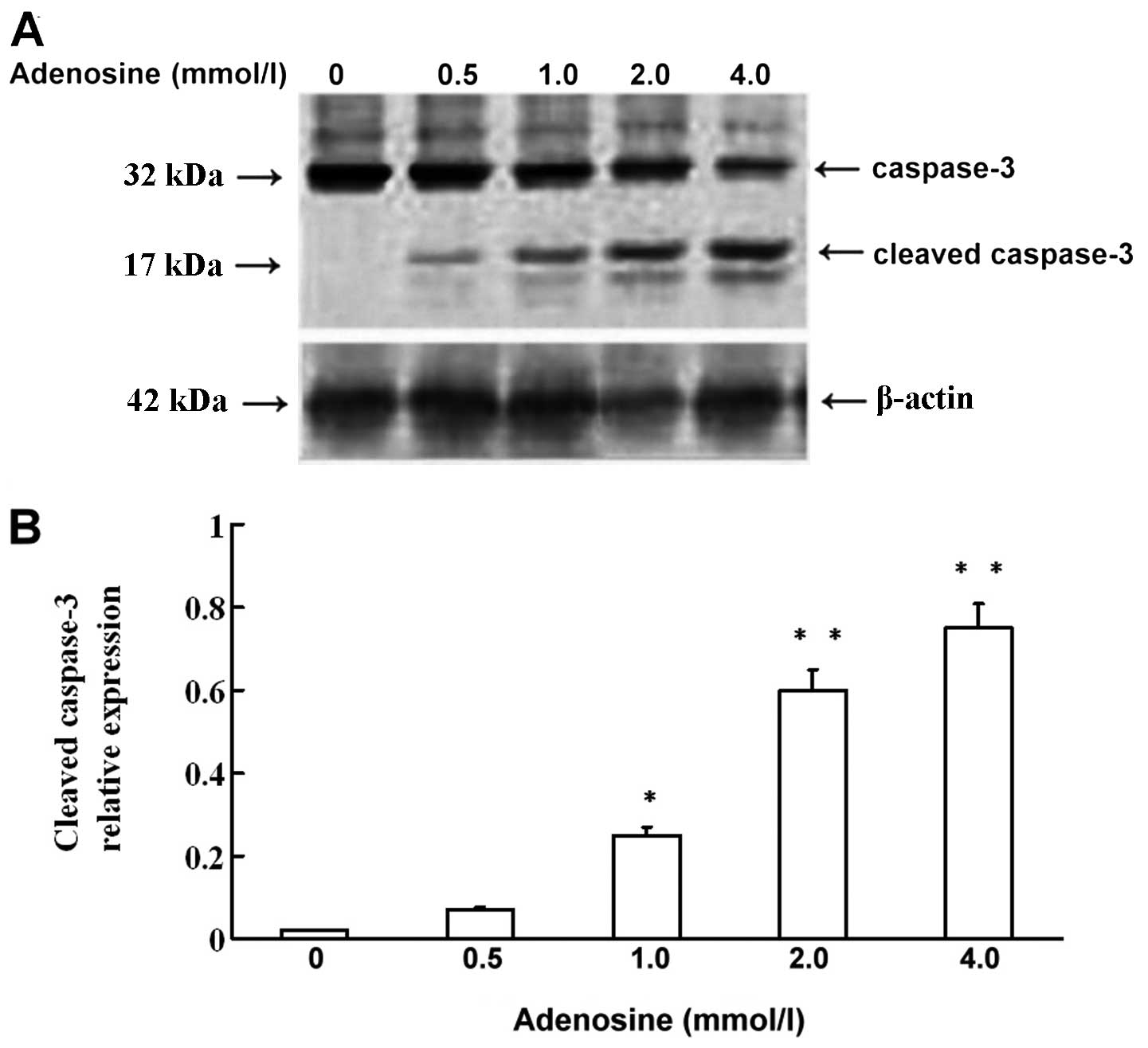
Apoptosis Induced By Adenosine Involves Endoplasmic Reticulum Stress In Ec109 Cells
Plos One Dissecting The Mechanisms Of Doxorubicin And Oxidative Stress Induced Cytotoxicity The Involvement Of Actin Cytoskeleton And Rock1
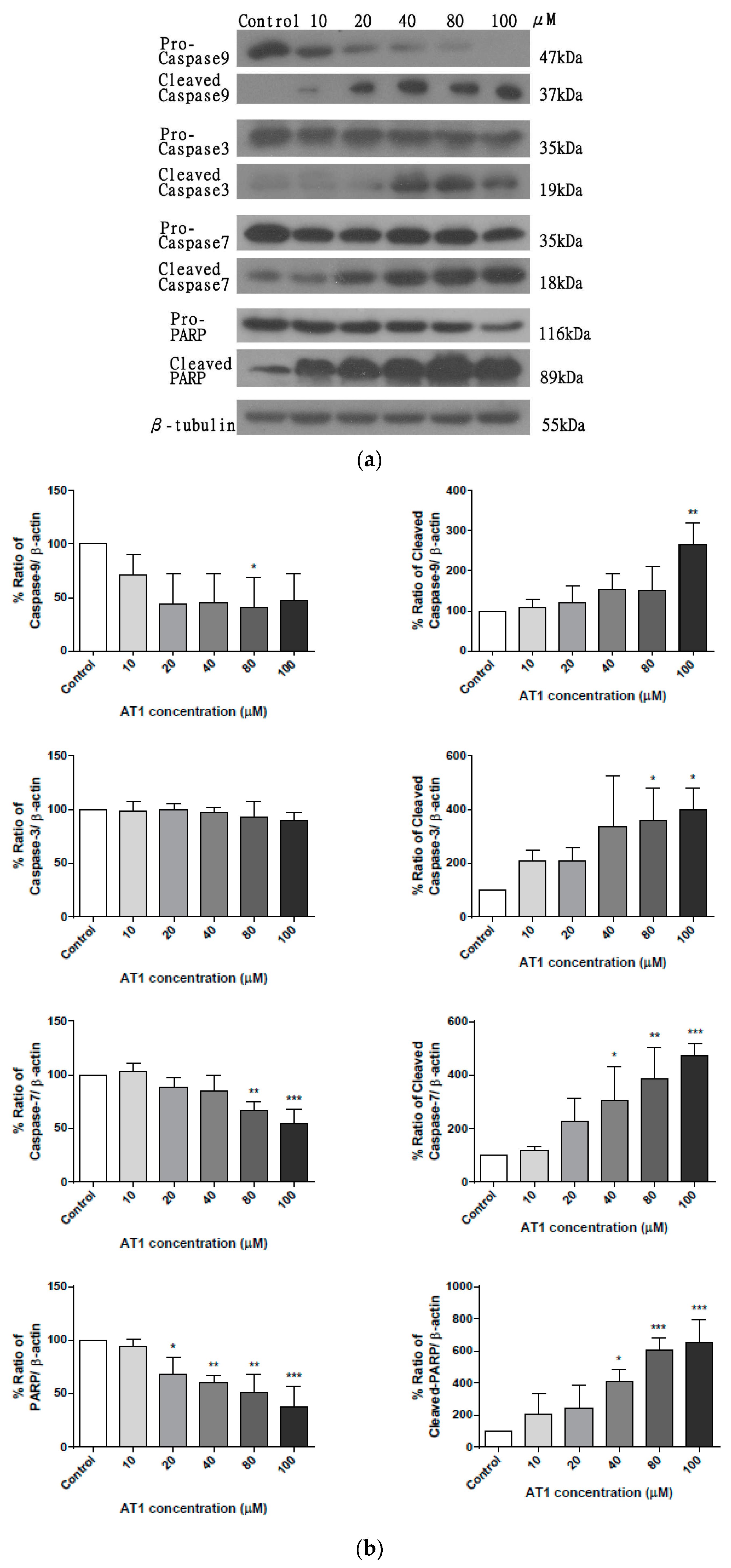
Molecules Free Full Text Anti Tumor Activity Of Atractylenolide I In Human Colon Adenocarcinoma In Vitro Html
Plos One Impact Of P53 Status On Trail Mediated Apoptotic And Non Apoptotic Signaling In Cancer Cells
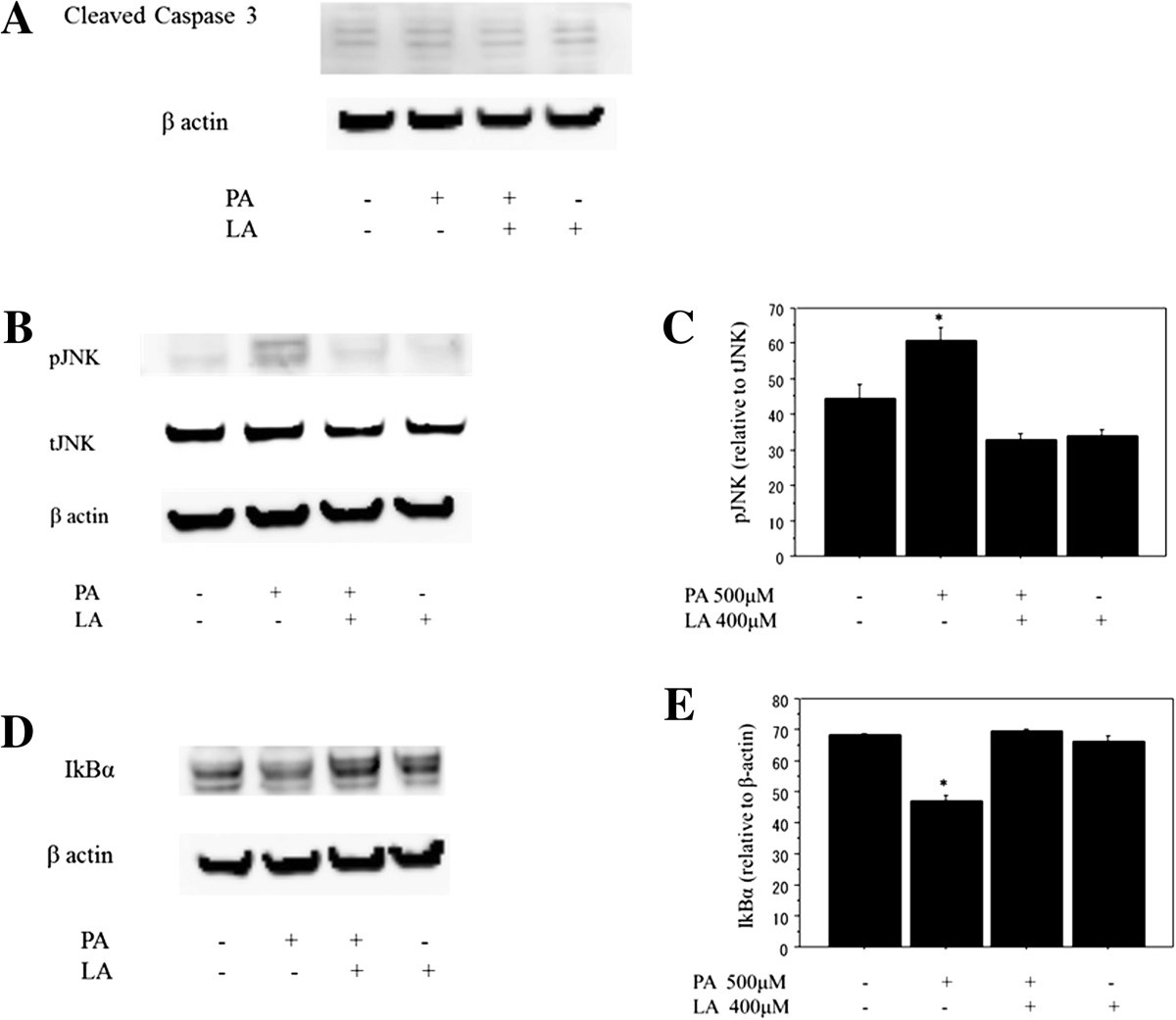
Linoleate Appears To Protect Against Palmitate Induced Inflammation In Huh7 Cells Lipids In Health And Disease Full Text

Western Blotting Shows That Co Treatment With The Caspase 8 Inhibitor Z Ietd Fmk Reverses Opg Induced Activation Of Caspase 8 And Caspase 3 In Ocs And Opcs

Caspase 3 Antibody
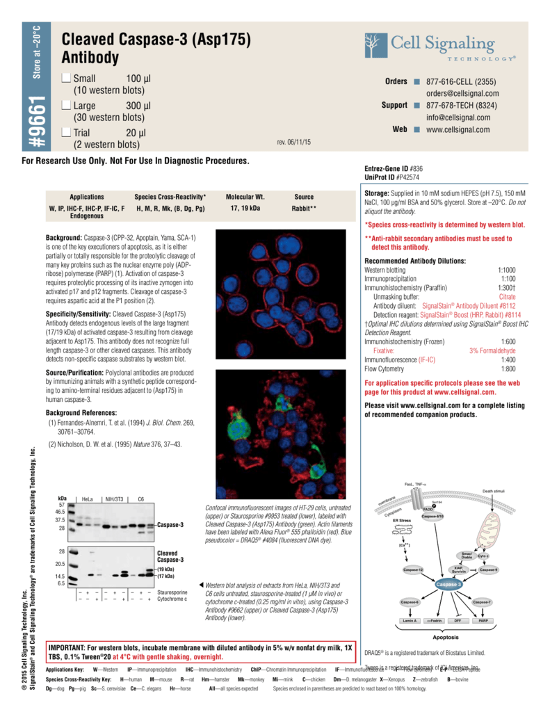
Cleaved Caspase 3 Asp175 Antibody

Western Blot And Densitometric Analysis Of Cleaved Caspase 3 Levels Download Scientific Diagram

Anti Casp3 Antibody Anti Human Active Caspase 3 Polyclonal Antibody Np 2
Cleaved Caspase 3 Asp175 5a1e Rabbit Mab 9664

View Image
The Effects Of Simvastatin On Hippocampal Caspase 3 And l 2 Expression Following Kainate Induced Seizures In Rats

Cleaved Caspase 3 P17 D175 Polyclonal Antibody Immunoway Biotechnology Company 抗体 病理抗体 诊断抗体

Sequential Application Of A Cytotoxic Nanoparticle And A Pi3k Inhibitor Enhances Antitumor Efficacy Cancer Research
Caspase 3 Control Cell Extracts
Caspase 3 P17 P19 Antibody 2 Ig Proteintech

Apoptosis Western Blot Cocktail Pro P17 Caspase 3 Cleaved Parp1 Muscle Actin Ab

Jci Ppar G Receptor Ligands Novel Therapy For Pituitary Adenomas

Western Blot Analysis Of Procaspase 3 And Cleaved Caspase 3 Panel A Download Scientific Diagram

Figure 2 From Inhibition Of Hsp 90 By Triptolide Tpl Augments Bortezomib Induced U 266 Cells Apoptosis Semantic Scholar
Plos One Vascular Endothelial Growth Factor Receptor 1 Inhibition Aggravates Diabetic Nephropathy Through Enos Signaling Pathway In Db Db Mice

Validated Anti Cleaved Caspase 3 P12 D175 Antibody Antibodyplus Antibody Trial And Validation

Caspase 3 Antibody Western C9598 Sigma Aldrich
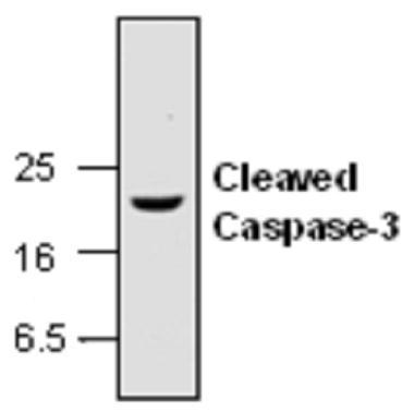
Anti Caspase 3 Active Antibody Gtx Genetex

Journal Of Acupuncture Research

Caspase 3 Activity Is Required For Skeletal Muscle Differentiation Pnas

Anti Casp3 Antibody Rabbit Cleaved Caspase 3 Polyclonal Antibody Np 2
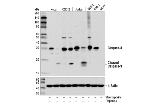
Caspase 3 Antibody D3r6y Cell Signaling Technology

Cleaved Caspase 3 Asp175 Antibody 9661s From Cell Signaling Technology Biocompare Com
Q Tbn 3aand9gcs2fe2gkdunlakufiorpmkspa Ijps Xeri1vavadfqfs7cdpoa Usqp Cau

Western Blot Assessing Cleaved Caspase 3 And Pro Caspase 3 Showed That Download Scientific Diagram


